![]()


![]()

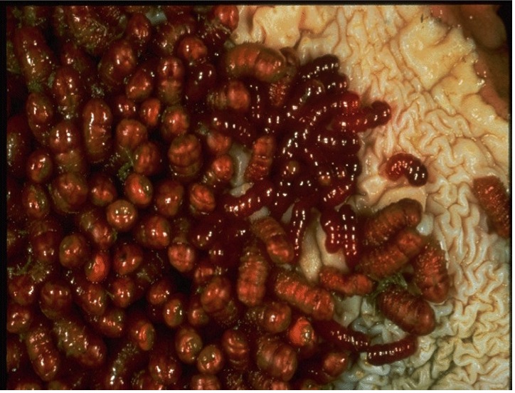
Stomach Bots are the larvae of Gasterophilus flies. The adult horse bot fly emerges a during the summer or fall season. After the fly emerges from the pupa, it quickly finds a mate, lays the eggs on the horse's coat and, on grooming, the eggs make their way to the mouth where they hatch in mouth as larvae. These larvae migrate to the stomach, causing an infestation for 8-10 months.
The larvae cause ulcerations in the lining of the stomach, and it is this which leads to disease. Disease is also caused by the larvae in mouth. Larvae here create borrowing holes, which in turn become infected. The 3rd stage larvae are passed in the faeces and over winters to develop the following summer.
Ascarids, (also known as roundworms) are parasites which are mainly a problem to foals. However, small numbers can also be carried by adults.
The adult worms are very large and can be up to 40cm in length. Once the larvae of this parasite are swallowed, they pass through the gut wall, via the liver to the lungs. Heavy infestations can cause coughing as the larvae travel through the lungs.
Adult worms can cause intestinal impaction. They pose a considerable threat to young horses and their developing immune systems, and they have to potential to kill by triggering colic. Even if a young horse escapes colic, a heavy ascarid burdened young horse will appear depressed and its normal growth will be affected.
The most common ascarid to infect horses is Parascaris equorum.The eggs laid by the large females pass out into pasture protected by a tough shell which equips them well for survival. They can withstand drying conditions and even freezing; waiting for that moment when a passing horse ingests them with grass.
Symptoms of Ascarid Infection In Foals
It goes without saying that the migratory habits of ascarids cause damage. However, the impact of ascarids in the horse will depend upon the degree of infection and the whether the larvae are in their migratory phase.
Ironically, this colic can be triggered by a dose of drenchan oral liquid dose. The worms are killed, fall away from the intestinal lining and cause a blockage. This is the primary reason that you not only need to drench orally dose young horses, but do so regularly to ensure there is no potentially fatal build-up of mature ascarids in the gut.
Symptoms of Ascarid Infection In Adult Horses
Adult horses are symptomless but can carry them and provide a reservoir for infestation to foals.
The common stomach worms of cattle are:
These stomach worms all have similar lifecycles and cause similar disease manifestations.
Ostertagia ostertagi (bimectin injection, bimectin pour on and bimectin plus)
OstertagiosisOstertagiasis, a parasitic gastritis caused by the nematode Ostertagia ostertagi, is the most important of the gastrointestinal helminthic infections of cattle in temperate climates. Disease caused by this brown stomach worm only occurs in first and sometimes second season grazing stock as immunity is developed over this period.
Life Cycle
Ostertagia has a direct life cycle typical of the other stomach worms.

Eggs passed in the faeces develop into first-stage larvae (L,), which hatch develop, and moult to become second-stage larvae (L2), which in turn develop and moult to the third infective stage (L3).
All of this occurs in the faeces pat, and the L3 then migrates under moist conditions onto the herbage. The L3 retains the outer sheath of the L3 and is the most resistant of the free-living stages.
Following ingestion, the parasitic cycle involves development through the L3 and L4 stages in the gastric glands. This usually takes 21 days, by which time the adult parasite emerges from the glands onto the surface of the abomasal mucosa.
The adults mate and the female worm will then produce thousands' of fertilised eggs which pass out of the animal in the faeces. The lifecycle of the larval stages depends on appropriate warm and moist conditions, which are found in the British Isles from May onwards. Under the appropriate conditions of temperature and humidity; the eggs will develop into larvae.
There are two presentations of OstertagiosisOstertagiasis and of Parasitic gastro-enteritis in cattle generally:
Type 1: Adult worms living in stomach attached to the lining, causing damage to the gut lining and deprive host of nutrients. Usually occurs in grazing season. Animals affected usually don’t thrive well and may have diarrhoea.
Type 2: Larval stage 4 burrows into stomach lining and emerge in large numbers causing explosive diarrhoea and may even be fatal. Usually occurs in December/January in the northern hemisphere following cold weather hibernation in the autumn but can occur in the autumn following warm weather hibernation in the summer.
Lungworm is also known as Hoose or Husk. Lungworm infestation is more prevalent in Europe than in North America.
 Courtesy of Department of Agriculture, Food and the Marine, Ireland
 Clinical signs of infection include coughing and difficulty in breathing. Affected cattle have an increased susceptibility to infective pneumonia. Immunity to lungworm develops quickly but is relatively short-lasting (approx. 6 months) in the absence of further infection.
 Courtesy of Department of Agriculture, Food and the Marine, Ireland
 At the start of each grazing season, following housing, cattle may have very little or no immunity to lungworm and thus are susceptible to new infections again. The periods of greatest challenge in temperate climates for animals are late summer and autumn. Reinfection Syndrome occurs in cows that are partly immune to Lungworm that are exposed to heavy larval challenge from pasture recently grazed by young susceptible animals. The cows may develop severe coughing and/ or milk drop as their immune system kills the migrating larvae. Such animals will not have Lungworm larvae detectable in faeces.
Life Cycle
The lungworm lifecycle is uniquely adapted amongst roundworms.

The life cycle of a lungworm begins with an ingestion of infective larvae. The larvae then penetrate the intestinal wall, and from here migrate into the lungs through the bloodstream. The infected larvae reside in the lungs until the development into an adult lungworm. The eggs of the adult hatch; producing L1 larvae. The eggs or L1 larvae that reside in the lungs are coughed up and then ingested back into the stomach and released into the environment via the faeces.
Lungworm and Fungus
The spread of lungworm is helped by a fungus (Pilobolus).
This orange fungus, regularly seen on cow dung pats in pastures, explodes, spreading the lungworm larvae all over the pasture.
Symptoms of Lungworm
How The Bimectin Range Can Help
Bimectin Injection, Bimectin Plus and Bimectin Pour On are all licensed to treat lungworm in cattle. Bimectin Injection is licensed to treat lungworm in cattle, sheep and pigs. Bimectin Paste is licensed to treat lungworm in horses.
| Product | Licensed for Lungworms in: | |||
|---|---|---|---|---|
| Cattle | Sheep | Pigs | Horses | |
| Bimectin Injection | Lungworms: (adult and fourth stage larvae)Dictyocaulus viviparus | Lungworms: Dictyocaulus filaria (adult and fourth stage larvae), Prostrongylus rufescens (adults) | Lungworms: Metastrongylus spp. (adults) | Not licensed for horses |
| Bimectin Plus Injection | Lungworms: (adult and fourth stage larvae)Dictyocaulus viviparous | Not licensed for sheep | Not licensed for pigs | Not licensed for horses |
| Bimectin Pour On | Lungworms: (adult and fourth stage larvae)Dictyocaulus viviparous | Not licensed for sheep | Not licensed for pigs | Not licensed for horses |
| Bimectin Paste | Not licensed for cattle | Not licensed for sheep | Not licensed for pigs | Lungworms: (adults and immatures) |
Louse populations are highest in cattle kept indoors during the winter months and heavy infestations cause irritation, leading to rubbing against feed barriers. This results in hair loss over the neck and shoulders and reduced DWG. Feed intake of cattle affected by external parasites such as lice, can drop by up to 10% and in cases of extreme infestation, anaemia may be seen.
Four species of lice infest cattle. They are classified as either biting lice (Damalina bovis) or sucking lice (Linognathus vituli or Haematopinus eurysternus, Solenopotes capillatus) . It not uncommon for cattle to be infected with more than one type of lice. This is particularly common in younger stock.
Life Cycle of Lice
The louse life cycle take 4 to 5 weeks and is similar for both sucking and biting lice. The eggs hatch and develop through 3 nymph stages to adults. This is illustrated below.
The thick, winter coat of the animal provide the ideal environment for development, providing a warm, humid and protected environment.
How Are Lice Transmitted?
As lice are usually transmitted by host contact, winter housing provides the ideal conditions for the transfer of lice between cattle.
There are various types of intestinal worms, which impact negatively on the health and productivity of animals. To find out more about some of the most common intestinal worms in the UK and Ireland, read on. Bimectin Injection, Bimectin Pour On and Bimectin Plus are all licensed as treatments for these intestinal parasites.
Cooperia spp
Several species of Cooperia are found in the small intestine of cattle; C punctata, C oncophora, and C pectinata are the most common and their life cycle is essentially the same as that of other trichostrongylids. Most of them are found in the first 10–20 ft. (3–6 m) of the small intestine and have a prepatent period is 12–15 days.
In heavy infections there is profuse diarrhoea, anorexia, and emaciation, but no anaemia; the upper small intestine shows marked congestion of the mucosa with small haemorrhages a milder form can occur but can be responsible for weight loss and poor productivity.
Hookworms (Bunostomum sp)
The pre-patent period is ∼2 months. Infection is by ingestion or skin penetration; the latter is more common.
Larval penetrating the lower limbs may cause uneasiness and stamping, particularly in housed cattle. Adult worms cause anaemia and rapid weight loss. Diarrhoea and constipation may alternate. Hypoproteinemic oedema may be present, but bottle jaw is rare.
The worms are readily seen in the first few feet of the small intestine, and the contents are often blood-stained. As few as 2,000 worms may cause death in calves. Local lesions, oedema, and scab formation may result from penetration of larvae into the skin of resistant calves.
Intestinal Threadworms (Strongyloides sp)
The intestinal threadworms, Strongyloides papillosus, are embedded in the mucosa of the upper small intestine. Infections are most common in young calves, particularly dairy stock. Although signs are rare, they may include intermittent diarrhoea, loss of appetite and weight, and sometimes blood and mucus in the faeces. Large numbers of worms in the intestine produce catarrhal enteritis with petechial and ecchymosis, especially in the duodenum and jejunum.
Nematodirus spp
Nematodirus helvetianus is generally recognized as the most common species in cattle, although other species, e.g., N spathiger and N battus, can also infect cattle. The eggs develop slowly; the infective third stage is reached within the egg in 2–4 week and may remain within the egg for several months. Eggs may accumulate on pastures and hatch in large numbers after rain to produce heavy infections over a short period. The eggs are highly resistant, and those passed by calves in one season may remain viable and infect calves the next season. After ingestion of infective larvae, the adult stage is reached in ∼3 weeks. Worms are most numerous 10–20 ft. (3–6 m) from the pylorus.
Oesophagostomum sp
These have a direct life cycle. The larvae penetrate primarily into the wall of the lower 10–20 ft. (3–6 m) of the small intestine but also into the cecum and colon, where they remain for 5–10 days and then return to the lumen as fourth-stage larvae. The pre-patent period in susceptible animals is ∼6 weeks. However, in subsequent reinfections, larvae become arrested for some time, and many never return to the lumen (host encystment).
Young animals suffer from the effects of adult worms, whereas in older animals, the effect of the nodules is more important. Infection causes anorexia; severe, constant, dark, persistent, fetid diarrhoea; weight loss; and death. In older, resistant animals, the nodules surrounding the larvae become caseated and calcified, thus decreasing the motility of the intestine. Stenosis or intussusception occasionally occurs. Nodules can be palpated per rectum, and the worms and nodules can be seen readily at necropsy.
Trichuris
Trichuris infections are common in young stock, but the numbers of worms are seldom large. Clinical signs are unlikely, but in occasional heavy infections, dark faeces, anaemia, and anorexia may be seen.
Mange in cattle is caused by mites. In the UK & Ireland there are 3 species of mange mites which affect cattle.
Mange mites cause irritation, hair loss, hide damage and a thickened, scaly skin. There are also associated with reduced productivity. Different mites affect different areas on the animal.
Timing
Mange is mainly a problem in autumn, winter and early spring as cattle are housed, with mites becoming less active and numbers reducing in the summer.
Chorioptic Mange (Chorioptes bovis)
Chorioptes bovis is common in housed cattle and is relatively harmless. It is more prevalent during the winter and often spontaneously regresses in summer. The pastern areas of the legs are preferred sites for the mites, though these mites are also found on the neck and tail head area. A high proportion of cattle can be infested without showing clinical signs. Some cattle will show hair loss which gradually increases in size and can cause irritation. Hide damage may be noted as cattle rub the affected areas.
Psoroptic Mange (Psoroptes bovis)
Psoroptic mange occurs worldwide and there are reports of disease in the UK caused by infestation with Psoroptes ovis thought to have arisen following the importation of infested cattle.
Clinical Presentation
Serum exudation and thickening of the skin particularly over the neck and over the dorsal midline is reported. In some cattle Psoroptic mange can cause severe clinical signs with adverse effects upon health and welfare. Bacterial infection of affected areas is common, leading to bleeding. If lesions are extensive, production losses will occur. In severe cases, death can result.
Sarcoptic Mange (Sarcoptes scabiei var.bovis)
Sarcoptic mange occurs worldwide but is rare in the UK & Ireland. Infestation causes severe pruritus with serum exudation and gross thickening of the skin, particularly over the neck. These mites are usually found on the neck and in the loin area next to the tail. As the name suggests, these mites borrow through the skin and cause severe irritation and skin damage. Skin will become thick and crusted and may become infected. This impairs productivity.
Sarcoptic mange can be transferred to human beings.
Adult warbles are circa 15 mm long, hairy, and bee-like in appearance. In late spring or early summer, they attach their eggs on cattle, particularly on the legs and lower body regions. The eggs hatch and penetrate the skin.
The first-stage larvae travel between muscles, along connective tissue, or along nerve pathways. They secrete proteolytic enzymes that facilitate their movement. During fall and winter, larvae migrate toward 2 different regions, depending on the species. H lineatum larvae migrate to the submucosal connective tissue of the oesophageal wall, where they accumulate for 2–4 months. H bovis larvae migrate to the region of the spinal canal, where they are found in the epidural fat between the dura mater and the periosteum for a similar period.
Beginning in early winter, the larvae arrive in the sub-dermal tissue of the back of the host where they make breathing holes through the skin. Cysts or warbles form around the larvae, which undergo 2 molts (second and third stage). The warble stage lasts 4–8 wk. Finally, third-stage larvae emerge through the breathing holes, drop to the ground, and pupate. Flies emerge from the pupae in 1–3 months, depending on weather conditions. Adult flies, which do not feed, live <1 week. The life cycle is complete in 1 year.
During periods of sunshine on warm days, cattle may run with their tails high in the air when chased by female warble flies, particularly H bovis.
Penetration of the skin by newly hatched larvae may produce a rash in older, previously infested cattle. The points of penetration are painful and inflamed and usually exude a yellow serum. Warbles may occur in the back from tail-head to the shoulders, and from top-line to about one-third the distance down the sides. Usually, the cysts are firm and raised considerably above the normal contour of the skin and, occasionally, develop in abscesses. The emergence of the grub, its forced expulsion, or its death within the cyst usually results in healing of the lesion without complications. Carcasses and hides of cattle infested with cattle grubs show marked evidence of the infestation and are reduced in value.
Withdrawal Period Milk:
N/A
Withdrawal Period Meat:
34 days
How The Bimectin Range Can Help

The Bimectin Horse Wormer (Ivermectin 1.87mg/g) is licensed to treat this condition in horses1. To find out more about the Bimectin Horse Wormer, click the image above.
1. Not licenced for hypobiotic larvae
How The Bimectin Range Can Help
The Bimectin Horse Wormer (Ivermectin 1.87mg/g) is licensed to treat this condition in horses1. To find out more about the Bimectin Horse Wormer, click the image above.
1. Not licenced for hypobiotic larvae
How The Bimectin Range Can Help

The Bimectin Horse Wormer (Ivermectin 1.87mg/g) is licensed to treat this condition in horses. To find out more about the Bimectin Horse Wormer, click the image above.
How The Bimectin Range Can Help

The Bimectin Horse Wormer (Ivermectin 1.87mg/g) is licensed to treat this condition in horses. To find out more about the Bimectin Horse Wormer, click the image above.


 Harmless Chorioptic mange