![]()


![]()

Los oxiuros son parásitos de color blanco-grisáceos con una cola larga que se va adelgazando hacia extremo distal. Las hembras de los oxiuros pueden alcanzar hasta 20 cm de longitud, mientras que los machos son significativamente más pequeños.
Aunque los oxiuros habitan en el intestino grueso y el colon, las hembras salen por las noches a ovopositar sobre el pelo alrededor de la grupa de los caballos, pudiendo poner hasta 60,000 huevos; después de esta acción, las hembras regresan al recto.
Las infestaciones por oxiuros generalmente se presentan en caballos de más de 18 meses de edad, aunque raramente llegan a causar problemas serios en los animales. A diferencia de otros parásitos, los oxiuros pueden afectar el sistema digestivo en los equinos, aunque no causan otros daños internos. Sin embargo, pueden provocar una irritación significativa.
En el momento que las hembras adultas liberan sus huevos sobre la piel, provocan una intensa irritación, obligando a los animales a restregarse sobre objetos duros, provocándose lesiones alrededor del ano. También se les puede observar mordiéndose y golpeándose sus cuartos traseros. No se detectan huevos durante el examen fecal; sin embargo se pueden observar huevos en la masa gelatinosa localizada alrededor del ano.
Dictyocaulus arnfieldi, es un nematodo que parasita los pulmones de los equinos, puede llegar a provocar tos y dificultad respiratoria, principalmente en las mañanas muy frescas o frías. Dicho parásito tiene una distribución mundial, siendo más frecuente en zonas con lluvias abundante. Estos nematodos son comunes en burros; en los que generalmente no representa un gran riesgo, pero que pueden diseminar huevos del parásito en los potreros. Debido a este punto, cuando caballos y burros pastan en los mismos sitios, los caballos fácilmente pueden infestarse y presentar signos cínicos de la parasitosis.
 Los parásitos adultos ovipositan en las vías respiratorias. Los huevos larvados (L1), junto con las secreciones, son transportadas a la faringe desde donde son expulsados al exterior por medio de la tos, o son ingeridos. Las L1 eclosionan durante su tránsito por el intestino y las larvas son expulsadas junto con las heces. En el ambiente, las L1 mudan en dos ocasiones, formando las L3 (fase infestante del parásito) en un lapso de una semana. Estas larvas habitan sobre hongos del género Pilobolus, frecuentes en las heces. Al explotar los esporangios del hongo, las larvas salen proyectadas hasta por 30 cm de distancia del excremento. Las L3 son sensibles a la sequedad, no obstante pueden hibernar si las condiciones son desfavorables. Los caballos se infestan al ingerir la L3. Una vez ingeridas, las larvas llegan al intestino, atraviesan la pared intestinal y, a través del torrente sanguíneo, llegan a los pulmones, se alojan en las vías respiratorias donde completan su desarrollo a adultos y se reproducen, provocando lesiones, irritación y obstrucción.
Una infestación masiva de Dictyocaulus arnfieldi causa irritación de la mucosa bronquial (bronquitis parasitaria), con producción abundante de moco, lo que causa dificultad respiratoria, tos severa y pérdida de apetito. La bronquitis se acompaña generalmente con neumonía crónica, edema pulmonar. Estas lesiones facilitan las infecciones bacterianas secundarias, que en presentaciones severas pueden llegar a causar la muerte, principalmente en potrillos.
El diagnostico se hace con base en los signos clínicos y la presencia de larvas (L1) en las heces, por medio de la técnica coproparasitoscopica de Baermann.
Los ascáridos, conocidos también como gusanos redondos, son parásitos que afectan principalmente a los animales jóvenes, aunque también se pueden encontrar en pequeñas cantidades en animales adultos.
Los parásitos adultos son grandes, pudiendo alcanzar hasta los 40 cm de longitud. Después de que las larvas de este parásito son ingeridas, cruzan la pared intestinal, pasan por el hígado hasta llegar a os pulmones. Una infestación severa puede provocar tos cuando las larvas migran por el parénquima pulmonar.
Los parásitos adultos pueden causar una impactación intestinal y constituyen una amenaza considerable para los caballos jóvenes y para el desarrollo de su sistema inmune; tienen un alto potencial de desencadenar problemas de cólicos. Aún cuando los animales jóvenes se recuperaran de los cólicos, una parasitosis severa los hará parecer deprimidos y será afectado negativamente su crecimiento.
El ascárido más común que afecta a los caballos es el Parascaris equorum. Los huevos liberados por las hembras; que salen con las heces, están protegidos por una pared gruesa que los hace resistentes al medio ambiente, proporcionándoles las condiciones para sobrevivir por largos periodos de tiempo. Pueden resistir desde condiciones de falta de humedad hasta el congelamiento, esperando que algún hospedero los ingiera junto con el forraje.
Signos de la Ascaridiasis en potrillos
No hace falta remarcar que los hábitos migratorios de estos parásitos, provocan daños al hospedero; sin embargo, el impacto de los ascáridos en los caballos dependerá del grado de infestación y de sí las larvas se encuentran en su fase migratoria.
Durante la fase migratoria de las larvas, los caballos pueden manifestar problemas respiratorios, pudiendo presentar descargas nasales y/o tos, en ocasiones acompañadas de fiebre, generalmente asociadas a infecciones bacterianas secundarias. La aplicación de antibióticos no mejora la signología en el animal, debido a que no tienen efecto contra los ascáridos.
Una carga parasitaria alta en los animales, podrá manifestarse con los signos clásicos de pelo hirsuto, pérdida de peso, abultamiento del abdomen y aletargamiento.
Los ascáridos representan un peligro notable; su tamaño es tal que en forma colectiva pueden llegar a bloquear el tracto intestinal de los animales jóvenes, pudiendo desencadenar la presentación de cólicos, que pueden llegar a ser fatales.
Irónicamente, la toma oral puede desencadenar estos cólicos, ya que, después de muertos, los parásitos son arrastrados en el intestino, pudiendo provocar bloqueos en el mismo. Esta es la razón principal por la que no solo es necesario desparasitar a los animales jóvenes, sino que se debe establecer una desparasitación rutinaria en todos los animales, con el fin de no permitir la presencia de parásitos adultos, con lo cual se evitará la presentación de cólicos fatales debido a esta parasitosis.
Signos de la Ascaridiasis en caballos adultos
Los caballos adultos no presentan signos manifiestos de la enfermedad, pero fungen como reservorios para la infestación de los potrillos.
While pinworms and roundworms have fairly efficient life cycles, strongyles have a very unusual method of reproduction, especially considering that they developed in a desert environment. Large and small strongyles share identical life cycles outside of the horse but behave very differently once they're ingested.
Eggs are passed in the fecal material, just like ascarid eggs. Unlike ascarid eggs, which remain in egg form until they are ingested by the horse, strongyle eggs hatch into larvae in the manure paddy when weather conditions are favorable. From here, they go through a series of three molts. It's the third stage that can infect the horse. Under optimal conditions, third-stage larvae can live for up to three months. For their species to survive, strongyles must be ingested shortly after molting to this stage.
The Typical Life Cycle of Small Strongyles
Strongyle Larvae in Dew Drop
 To increase its chances for survival, the larva climbs up on blades of grass where a horse can ingest it while grazing. This is less likely under natural conditions, because the horse herd that deposited this larvae-ridden manure may have migrated many miles.
Strongyles are prolific egg layers. A single horse can pass 75-100 million eggs daily. The eggs can crawl up and down a blade of grass many times or bury themselves in the soil to protect against adverse weather.
Small Strongyles
Small strongyles also developed another unique trait - the ability to encyst themselves in the horse's tissues and remain in an encysted state for a prolonged time. Unfortunately, it causes real problems for the horse.
This encysted state helps the small strongyle: When larvae are ingested, they immediately penetrate the bowel lining and begin a two- to three-month migration in the intestinal tract. The immune system recognizes these migrating parasites as foreign invaders and sends its defense cells to deal with the invasion. Because the migrating parasites are too large and are moving, the defense mechanism can't deal with them effectively. The only result from the immune response is mucosal inflammation.
If the horse already has an adult population of this parasite, there appears to be a communication between the adult population and the migrating larvae. This negative feedback tells the larvae, "We have enough adults laying eggs in the intestine. Slow your migration and wait your turn to become an adult." Once the larva slows its migration, the immune system can deal with this invader and surround it with scar tissue.
Cycle Requires Only Occasional Exposure
The small strongyle has developed a way to make the horse an egg-laying machine, and keep it that way with only occasional exposure to the larvae. Horses only have to pick up these infected larvae one time every two or three years for the small strongyle's life cycle to function.
Small strongyles, however, are the most significant causes of chronic under-performance, loss of condition, feed inefficiency and predisposition to secondary disease.
Strongylus vulgaris Migration
 The pattern of migration for Strongylus vulgaris, a large strongyle, is quite different. Eggs are passed in the feces and develop through two larval stages. When a third-stage larva is ingested, it migrates to the large intestine, where it loses its sheath and penetrates the intestinal wall. The fourth-stage larvae penetrate the submucosal arterioles and migrate to the cranial mesenteric artery - the artery that supplies blood throughout the intestinal tract - where they molt to the fifth stage. These larvae return via intestinal arteries to the gut, where they reproduce.
Arterial Damage From Large Strongyles
No matter where a strongyle larva penetrates, leaves the gut and begins its migration, it will always end up at the same spot - the beginning of the cranial mesenteric artery, which is the primary blood source for the intestinal tract. All these larvae in one spot cause tremendous damage and reaction. In fact, in rare cases, the artery can rupture, causing rapid death. The more common problem is a weakening of the arterial wall, which can lead to a malformed artery - an aneurysm. This malformation causes abnormal blood flow, which can lead to formation of blood clots in the artery. These clots cling to the artery walls like clusters of grapes. Should one of these clots break free, it will be forced downstream in the blood supply of the intestines, where it may block blood flow. This situation, called thromboembolic colic, can result in serious illness and death.
The pinworm is a grey/white worm with a long tail, which tapers to a point. While the male is significantly smaller, the female pinworm can reach up to 20cm in length.
Infective pinworm eggs are ingested orally and, once in the colon, the larvae develop through various stages before becoming sexually mature in about five months. As horses migrate, they take the eggs and adults with them.
Pinworms have the most efficient life cycle of all the parasites that infect the horse. They don't migrate through any organ tissue, and they have developed a means of reproduction by which the eggs don't leave the herd of horses.
Although pinworms live in the large intestine and colon, the adult worm emerges at night to lay eggs on the skin around the rump. After laying her eggs, the female pinworm will then return inside the rectum. A female pinworm can lay up to 60,000 eggs per day.
Pinworm infestations commonly occur in horses older than 18 months, but rarely cause the animal major problems. Unlike many other worms, the pinworm will not cause damage to the horse’s digestive system, nor will it cause other internal damage. However, they will cause significant irritation.
When the adult worm lays her eggs it can cause intense irritation leading to rubbing and self-harm around the anus. Biting and licking of hindquarters may also be observed. There are no eggs on faecal examination. However, eggs may be seen in a gelatinous mass around the anus.
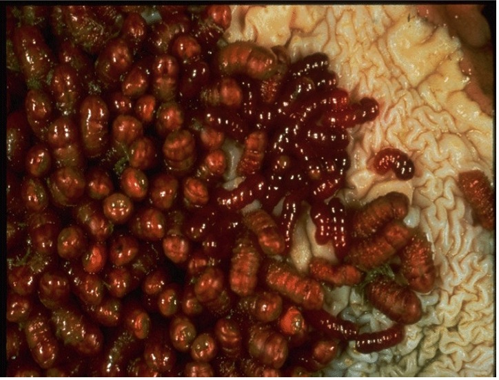
Stomach Bots are the larvae of Gasterophilus flies. The adult horse bot fly emerges a during the summer or fall season. After the fly emerges from the pupa, it quickly finds a mate, lays the eggs on the horse's coat and, on grooming, the eggs make their way to the mouth where they hatch in mouth as larvae. These larvae migrate to the stomach, causing an infestation for 8-10 months.
The larvae cause ulcerations in the lining of the stomach, and it is this which leads to disease. Disease is also caused by the larvae in mouth. Larvae here create borrowing holes, which in turn become infected. The 3rd stage larvae are passed in the faeces and over winters to develop the following summer.
Here is more information about bots and detail regarding how the Bimectin Range can be an effective weapon in your bot treatment strategy.
The Bot: More Than a Pest
Bots Flies are common in most stables. Often swatted at, but rarely hit, they are a pest poorly tolerated by horse and owner. Bot flies can be much more than just pests, however. The annoyance and distraction they cause can interfere with feeding and affect nutrition. The migration of bot larvae under the skin in mucous membranes causes lesions that may provide openings for infection. Flies also carry diseases that can seriously harm your horse's health and performance. Without treatment, bots can cause severe damage in the stomach and intestine of your horse.
A Long Life Cycle
Adult bot flies are brown, hairy and bee-like, with one pair of wings, and measure about 3/4". The bot larva is also 3/4" long, with a narrow, hooked end and a broad, rounded body. In the summer months, adult bot flies are a common sight around horses. Yet this adult stage is just a brief part of the bot fly life cycle. Female bot flies have no mouth parts, so they cannot feed. They live on stored reserves only long enough to lay eggs on the hair around a horse's eyes, mouth, nose, or on the legs. Moisture from the skin or from the horse's licking causes the eggs to hatch into larvae.
The Bot Life Cycle
After a three-week developmental period in the mouth, bot fly larvae of both species, Gasterophilus intestinalis and Gasterophilus nasalis, migrate and attach themselves to the mucus lining of the horse's stomach and remain there during the winter. After about 10 months, they detach from the lining and are passed out of the body through the feces. The larvae burrow into the ground and mature. Depending on the conditions, adults emerge in three to 10 weeks. Adult females deposit eggs on the horse's legs, shoulders, chin, throat and lips. Depending on geographic location, the life cycle of bot flies is not fixed to only certain times of the year, and bot larvae can be active in horses anywhere from August to May.
Egg laying begins in early summer. Eggs of the two species differ in color and placement.
G. intestinalis – G. nasalis
G. intestinalis lays up to 1,000 pale-yellow eggs on the horse's forelegs and shoulders. Moisture and friction from the horse's licking itself cause the eggs to hatch in about seven days. After hatching, G. intestinalis larvae are licked into the mouth.
G. nasalis lays about 500 yellow eggs around the chin and throat of the horse. These eggs are not dependent on the horse's licking them to hatch. G. nasalis burrows under the skin to the mouth, wandering through it for about a month before migrating to the stomach for overwintering. Then the cycle begins again.
Signs of Bot Infestation
Horses that show no outward signs of illness can be severely infested, giving no clue to damage occurring inside. However, some horses do show signs of infestation, including an inflamed mouth area and stomach irritation. Infestation with bot larvae may cause ulcers in the stomach lining. If the infestation is severe, the opening from the stomach to the intestines may be blocked, which can cause irritation, ulcers and even colic. The burrowing larvae can cause small tears in the skin, which can become infected.
Treatment for Bots
Traditionally, horses are treated for bots in the fall, after a frost that kills the adult flies, and again in the spring, to rid the stomach of all the larvae. In the past, the treatment was worse than the disease, with extremely toxic chemicals given via stomach tube to the horse. Modern anthelmintics like Ivermectin, the active ingredient in Bimectin Paste 1.87%, are extremely effective and safe in the treatment of bots and have had an impact on lowering the number of bot flies in areas where good anthelmintic treatment is practiced.
There are three major species of Large Strongyles, which are Strongylus vulgaris, S edentates, and S equinus. Of these, the most common is Strongylus vulgaris.
Horses become infected through the ingestion of infective larvae, which ex-sheath in the intestine and migrate, before developing to maturity in the large intestine.
Large Strongyles are blood feeders and they ingest mucosal plugs as they move through the intestine. Anaemia can result from this blood loss. Large Strongyles also cause weakness, loss of weight and diarrhoea. As a result of damage to mesenteric artery, Large Strongyles can also cause colic.
Large Strongyles can be extremely dangerous to the horse. However, low resistance levels mean that control is relatively straightforward.
Lungworm can cause coughing and respiratory difficulty in horses, although they are relatively uncommon. These worms are common in donkeys and easily transfer to horses grazing the same pasture.
Horses become infected by eating the worm larvae from the pasture. The larvae will then move to the lungs, where they become adult. These adults, living in the airways, can cause obstruction leading to coughing and breathlessness. The adults lay eggs which are coughed up and swallowed to then be passed out in the droppings; thus continuing the cycle.
Diagnosis usually takes place on the basis of clinical symptoms and the presences of eggs in faeces.
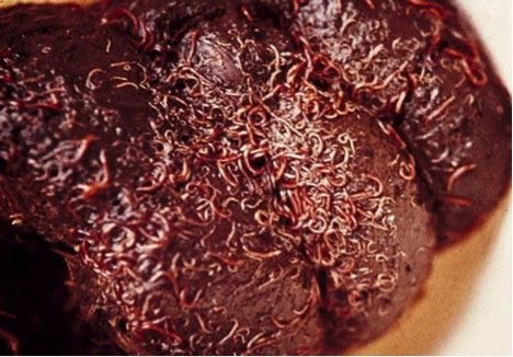
Small Strongyles are the most common equine worm, and the adult worms alone cause little ill effect.
It is the hypobiotic larvae emerging from gut wall which cause disease. Once ingested, the small larvae have the ability to burrow into the lining of the intestine. When they emerge, they can do so in very large numbers, causing severe intestinal damage and diarrhoea.
Treatment using ivermectins can control the adult and immature stages of this parasite and will minimise the numbers eggs being shed onto the pasture. The larvae are difficult to treat and resistance to wormers is a major issue. Pasture sweeping, wormer rotation, FECRTs and paddock rotation are all good ways to minimise risk.
The Typical Life Cycle of Small Strongyles
The pinworm is a grey/white worm with a long tail, which tapers to a point. While the male is significantly smaller, the female pinworm can reach up to 20cm in length.
Although pinworms live in the large intestine and colon, the adult worm emerges at night to lay eggs on the skin around the rump. After laying her eggs, the female pinworm will then return inside the rectum. A female pinworm can lay up to 60,000 eggs per day.
Pinworm infestations commonly occur in horses older than 18 months, but rarely cause the animal major problems. Unlike many other worms, the pinworm will cause damage to the horse's digestive system, nor will it cause other internal damage. However, they will cause significant irritation.
When the adult worm lays her eggs it can cause intense irritation leading to rubbing and self-harm around the anus. Biting and licking of hindquarters may also be observed. There are no eggs on faecal examination. However, eggs may be seen in a gelatinous mass around the anus.
CÓMO LA LÍNEA BIMECTIN® PUEDE AYUDAR
Bimectin Injection Injection está autorizado para el tratamiento, prevención y control de una amplia de variedad de parásitos internos y externos que afectan a los cerdos. Para obtener mayor información sobre Bimectin Injection, y su uso, haga clic aquí.